Conditions Retinal detachment
ICD codes: H33 What are ICD codes?
Retinal detachment is a rare severe eye condition. It can lead to vision loss if left untreated. Light flashes, dark specks or a black shadow in the field of vision are typical signs. Vision damage can often be avoided if retinal detachment is treated promptly.
At a glance
- Retinal detachment is a rare eye condition, which can have a severe impact on a person’s sight.
- In this process, the retina detaches from the inner surface of the eyeball and is damaged as a result.
- Light flashes, dark specks or streaks in the field of vision are typical symptoms.
- Urgent medical treatment is needed if these symptoms occur.
- If a retinal detachment is detected and operated on early, vision loss is often avoided.
Note: The information in this article cannot and should not replace a medical consultation and must not be used for self-diagnosis or treatment.


What is retinal detachment?
The retina lines the eyeball from the inside. It contains millions of sensory cells responsible for perceiving color and light. However, if small tears or holes develop in the retina, they can detach from the underlying layer.
A retinal detachment causes a variety of vision problems, such as flashes of light, floaters, black dots, shadows in the field of vision, or loss of vision in one eye.
It is important to see an ophthalmologist as soon as possible if such signs appear. Retinal detachment is an emergency and must be treated as soon as possible. Otherwise, the retina can detach further, which can lead to blindness in the affected eye.

Important: Retinal detachment is an emergency that requires immediate treatment.
What are the symptoms of retinal detachment?
Retinal detachment is painless. People with retinal detachment perceive flashes of light, even in the dark and sometimes with their eyes closed. Some people see floaters, rings, or black dots before their eyes, while others see “soot spots” or a “shower of black spots” – meaning a large number of black dots that can look like a spiderweb. Some may see a large black area that moves downwards like a curtain falling or makes their vision cloudy.
These symptoms may be accompanied by floaters (sometimes called “mouches volantes” – French for “flying flies”), although these may also occur with other eye conditions. Floaters appear as streaks and dots not unlike a swarm of small flies. This symptom may be harmless and occur temporarily due to vitreous opacities. However, it is advisable to consult a doctor if floaters remain in the field of vision for a long time.
The symptoms that occur depend, among other things, on the location where the retina detaches. If the center of the retina (macula) is affected, vision is blurred or distorted.

Important: Seeing black shadows, “curtains” coming down, or flashes of light are warning signs of retinal detachment. It is important to see a doctor immediately in this case.
What causes retinal detachment?
The retina is made up of two layers – the upper, neural layer contains photoreceptors and nerve cells. Below this is the pigment epithelium, or pigment layer. The pigment layer draws the nerve layer in through a thin film of fluid and supplies it with nutrients.
If, for example, the gel-like fluid from the center of the eye penetrates between the two layers through a tear, they separate from each other. As a result, the photoreceptors and nerve cells no longer receive an adequate supply of blood. This results in the typical visual impairments described.
Not every retinal detachment is caused by tears. Sometimes scarring on the retina or fluid accumulation resulting from other conditions is the cause – though both are significantly less common.
What factors increase the risk of retinal detachment?
The risk of experiencing retinal detachment increases with age.
People who have moderate to severe myopia (short-sightedness) are also at an increased risk of retinal detachment because their eyeball is slightly too long. As a result, the retina is always under a little tension and can tear more easily.
The risk of retinal detachment is also increased if retinal detachments run in the family.
Anyone who has ever undergone eye surgery or has injured their eye, for example during sports, also has a higher risk of retinal detachment.
Retinal detachment occurs more frequently in people with diabetes because diabetes can lead to retinal damage.

How common is retinal detachment?
Retinal detachment is rare. Experts estimate that in Central Europe, retinal detachment occurs in approximately 10 to 25 out of every 100,000 people each year.
The condition becomes more common with age. By the age of 85, about 3 percent of all people develop a retinal detachment. Men are affected more often than women. Children and people under the age of 30 are, however, only rarely affected.

What is the outlook for people who experience retinal detachment?
If the retina has detached, rapid diagnosis and surgery are crucial to maintain or restore eyesight. Without surgery, an individual can go permanently blind in the affected eye.
With a severely torn retina, it may become completely detached within 1 to 2 days. However, if the tears and holes are small, the retina detaches over a longer period of weeks or even months.
What can be done to prevent vision loss due to retinal detachment?
Age-related retinal detachment often cannot be prevented by means of preventive measures. However, it is important to have changes in the field of vision examined promptly by an ophthalmologist. This is because if a retinal detachment is detected and treated early, this can prevent permanent vision loss.
If there is a higher risk of retinal detachment – for example due to shortsightedness – regular check-ups are important. To prevent a detachment of the retina, early precursors like retinal tears can be treated in an eye doctor’s office.
Two methods are possible: laser treatment (thermal laser coagulation) or cold therapy (cryocoagulation). In this process, heat or cold is used to reattach the retina around the tear or hole to the underlaying layer. In this way, tears remain but cannot grow larger.
The risk of eye injury and resulting retinal detachment is reduced by the individual wearing safety goggles for certain activities. These include tasks such as sawing, drilling, or hammering – but also contact and ball sports, such as hockey or squash.
How is retinal detachment diagnosed?
The ophthalmologist starts by asking about symptoms, other conditions or injuries. Then, an eye test is often performed first to check visual acuity. To examine the retina at the back of the eye in greater detail, a fundus examination (ophthalmoscopy) is performed.
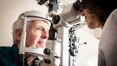
For the examination, eye drops are first administered to dilate the pupil. Using a combination of a microscope and a special type of light called a slit lamp, the ophthalmologist can examine the back of the eye to detect changes to the retina. Tears, holes, creases or bleeding indicate retinal detachment. After the examination, you will be temporarily sensitive to light and your vision will be somewhat blurred.
Sometimes, an ultrasound scan is also needed to produce a definitive diagnosis.
How is retinal detachment treated?
The sooner a retinal detachment is treated, the better. In most cases, the retina can be reattached. Sometimes, several interventions are necessary for this. How well you can see afterwards depends, among other things, on where the retina has detached and how extensive the damage already was.
If the ophthalmologist sees during the examination that there is a tear in the retina but that the retina has not yet become detached, cryotherapy or laser treatment may be sufficient. Using a special laser or freezing probe, the upper layer of the retina can be re-attached to the lower layer. The laser beam or freezing gas is applied to the retina, causing scarring that knits the two layers back together.
If the retina has already detached, it can be re-attached using surgery. Various surgical procedures can be used, depending on the type and severity of the retinal detachment that has occurred.
- Scleral buckling surgery: This involves using a silicone buckle or an encircling band to indent the eyeball from the outside, so that the retina rests against the underlying layer once more.
- Removal of the vitreous body: In this procedure, the surgeon removes the gel-like fluid of the vitreous body and replaces it with gas or a silicone oil to apply pressure to the retina. This presses the two layers back together. After some time, the gas is naturally replaced by the body’s own fluid. To remove the silicone oil, however, a further procedure is necessary.
In most cases, the detached retina can be reattached surgically. This often improves vision in this area as well. However, if the center of the retina (macula) is affected, vision usually remains permanently impaired – vision is blurred and has less contrast.

Important: Surgery on the retina should be performed in a specialized hospital or center.
- American Optometric Association. Care of the Patient with Retinal Detachment And Related Peripheral Vitreoretinal Disease. 2004.
- Bechrakis NE, Dimmer A. Rhegmatogene Netzhautablösung Epidemiologie und Risikofaktoren. Ophthalmologe 2018; 115: 163–178.
- D'Amico DJ. Clinical practice. Primary retinal detachment. N Engl J Med 2008; 359(22): 2346-2354.
- Deutsche Ophthalmologische Gesellschaft (DOG), Retinologische Gesellschaft (RG), Berufsverband der Augenärzte Deutschlands (BVA). Risikofaktoren und Prophylaxe der rhegmatogenen Netzhautablösung bei Erwachsenen (S1-Leitlinie). AWMF-Registernr.: 045-025. 2022.
- Feltgen N, Walter P. Rhegmatogenous retinal detachment - an ophthalmologic emergency. Dtsch Arztebl Int 2014; 111(1-2): 12-21; quiz 22.
- Gelston CD, Deitz GA. Eye Emergencies. Am Fam Physician 2020; 102(9): 539-545.
- Kang HK, Luff AJ. Management of retinal detachment: a guide for non-ophthalmologists. BMJ 2008; 336(7655): 1235-1240.
- Lang GK. Augenheilkunde. Stuttgart: Thieme; 2014.
- Sena DF, Kilian R, Liu SH et al. Pneumatic retinopexy versus scleral buckle for repairing simple rhegmatogenous retinal detachments. Cochrane Database Syst Rev 2021; (11): CD008350.
- Ullrich M, Zwickl H, Findl O. Incidence of rhegmatogenous retinal detachment in myopic phakic eyes. J Cataract Refract Surg 2021; 47(4): 533-541.