Conditions Cauliflower ear
ICD codes: M95.1 What are ICD codes?
Blunt trauma to the ear can cause bruising of the auricle/pinna (external ear) between the cartilage and the layer of connective tissue around it (the perichondrium). The cartilage shrivels up, folds in on itself and forms fibrous, bumpy structures that appear similar to a piece of cauliflower. Cauliflower ear can be prevented from developing if the bruising is treated early.
At a glance
- Cauliflower ear is a permanent deformity of the external ear.
- It occurs as a result of untreated bruising caused by blunt trauma to the ear.
- The injured tissue shrivels up, folds in on itself and forms new fibrous cartilage, giving the external ear an appearance similar to a piece of cauliflower.
- People who practice martial arts or play contact sports are at a particularly high risk of developing cauliflower ear.
- Cauliflower ear is prevented if the bruising is treated quickly.
Note: The information in this article cannot and should not replace a medical consultation and must not be used for self-diagnosis or treatment.
")
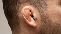
What is cauliflower ear?
Cauliflower ear is a permanent deformity of the external ear. It occurs when the external ear (auricle/pinna) is injured – by an impact, for example – and a bruise (hematoma) forms.
If this bruising is left untreated, the cartilage in the outer ear can no longer receive an adequate supply of nutrients. As a result, it becomes deformed and some parts may even die. In addition, the hematoma is replaced firstly by soft connective tissue and later by cartilage. This leads to typical changes in the auricle/pinna, which cause its surface to appear bumpy and lumpy – similar to a piece of cauliflower.
Bruising of the ear is particularly likely to occur as a result of failing to wear headgear when playing contact sports or practicing martial arts.

Cauliflower ear is usually a cosmetic problem. Some people find the deformity of their external ear distressing. Scarring is rare. Complications such as infection or tearing of the auricle are also rare.
What are the symptoms of cauliflower ear?
The most striking feature of cauliflower ear are the gnarled growths of tissue in the external ear, which make it appear swollen and deformed.
Other possible symptoms are:
- pain
- reddish or blueish discoloration
- swelling due to fluid retention (edema)
How does cauliflower ear develop?
Cauliflower ear usually develops as a result of a forceful impact to the ear. It frequently occurs in people who practice martial arts and play contact sports, in particular rugby, wrestling and boxing.
The connective tissue surrounding the cartilage is injured. As a result, blood pools between the cartilage and the layer of connective tissue around it (the perichondrium), leading to a bruise (hematoma). When this occurs, the cartilage in the area no longer receives an adequate supply of fresh blood and some parts of it may die.
If this type of bruising is left untreated, it will gradually be replaced firstly by connective tissue and later by newly formed cartilage. As a result, the external ear looks swollen and takes on an appearance similar to that of a piece of cauliflower.
How is cauliflower ear diagnosed?
If a person with suspected cauliflower ear consults their family doctor, the doctor will first take their medical history and ask whether they have recently had an accident or a bang on the ear. The doctor will then examine the ear and external ear for any swelling or discoloration.
Further tests, such as an ultrasound or computed tomography (CT) scan may be considered in order to exclude other injuries.
How is cauliflower ear treated?
Cauliflower ear is prevented by rapid treatment of bruising to the external ear.

To prevent the bruising from spreading, it is important to immediately cool the area and put pressure on it, for example using a compression bandage.
Painkillers can alleviate acute pain.
Doctors can drain excess blood from small hematomas using a needle. This is done under local anesthetic.
If the hematoma is larger or has been developing for more than two days, surgical intervention is often required. The procedure involves the doctor removing fluid and clotted blood under local anesthetic.
This allows the connective tissue (perichondrium) to re-attach to the underlying cartilage so that the blood vessels in this tissue can once again supply the cartilage with fresh blood. A compression bandage is used for support. It prevents blood from accumulating between the cartilage and the connective tissue once again and creating a new hematoma.
- DynaMed (Internet), Ipswich (MA). Auricular Hematoma – Emergency Management. EBSCO Information Services. Record No. T917237. 2018 (1995). Aufgerufen am 02.07.2021.
- Krogmann RJ, Jamal Z, King KC. Auricular Hematoma. [Updated 2021 Jan 20]. In: StatPearls [Internet]. Treasure Island (FL): StatPearls Publishing; 2021 Jan-. Aufgerufen am 02.07.2021.
- Patel BC, Skidmore K, Hutchison J, et al. Cauliflower Ear. [Updated 2021 Feb 25]. In: StatPearls [Internet]. Treasure Island (FL): StatPearls Publishing; 2021 Jan-. Aufgerufen am 02.07.2021.
- UpToDate (Internet). Assessment and management of auricular hematoma and cauliflower ear. Wolters Kluwer 2020. Aufgerufen am 02.07.2021.
In cooperation with the Institute for Quality and Efficiency in Health Care (Institut für Qualität und Wirtschaftlichkeit im Gesundheitswesen – IQWiG).
As at: