Conditions Macular degeneration
ICD codes: H35.3 What are ICD codes?
Eyesight slowly deteriorates for most people as they age. Diseases such as age-related macular degeneration cause further deterioration. Objects appear blurry and distorted when looked at directly. Reading also becomes more difficult.
At a glance
- Age-related macular degeneration (AMD) is a chronic disease that is triggered by a metabolic disorder. Typically, both eyes are affected.
- Macular degeneration impacts the eyesight in terms of visual acuity.
- It occurs more often in older people.
- Age-related macular degeneration typically does not lead to total blindness. While around 1 percent of people between 65 and 75 years of age have AMD, the condition affects 10 to 20 percent of women and men over 85.
- Age-related macular degeneration typically does not lead to total blindness.
Note: The information in this article cannot and should not replace a medical consultation and must not be used for self-diagnosis or treatment.

What is macular degeneration?
The eyesight of a person deteriorates with age. However, diseases can also contribute to the further deterioration of a person’s eyesight, sometimes to the point of blindness.
Age-related macular degeneration (AMD) also causes advanced visual impairment.
AMD is a chronic disease that is triggered by a metabolic disorder. Typically, both eyes are affected.
AMD originates in the macula. The macula is the part of the retina that is particularly important for visual acuity. Most often, eyesight is only impaired in the advanced stage of the disease.
There are two types of age-related macular degeneration: wet and dry. The former causes visual impairment more quickly. There is no cure for either type.
Treatment of the wet form of AMD can be slightly successful though. Visual acuity can be preserved and sometimes even improved. Treatment can slow down the progression of the disease.
What are the symptoms of macular degeneration?
Age-related macular degeneration (AMD) causes visual acuity (for which the macula is responsible) to slowly deteriorate. The macula, also called the macula lutea or yellow spot, is located at the center of the retina of the eye. It helps a person read, drive a car, or recognize faces.

A central vision disorder can cause objects a person focuses on directly to appear blurry and distorted.
Those with advanced AMD can no longer see objects. Objects at the edge of the field of view can typically still be seen but not recognized well.
What causes macular degeneration?
The metabolism in the retina is impaired in people with age-related macular degeneration.
Waste products, which are typically broken down by the body, are produced during the metabolic process. When they are not broken down properly, deposits form. These “drusen” – yellow deposits under the retina – prevent the retina from being properly nourished.
The effects vary based on the type of AMD in question:
- With dry AMD, the light-sensitive cells of the retina are slowly destroyed. Furthermore, pigment changes below the retina can occur.
- With wet AMD, the body reacts to the deposits by forming new blood vessels below the retina. If they become permeable, blood and liquid enter the retina and damage the cells. Moreover, the new blood vessels can raise the retina.
People with a close relative who has been diagnosed with AMD are at a slightly higher risk of also developing AMD. In addition, smokers are more likely to develop AMD, and at an earlier age.
How common is macular degeneration?
The majority of people develop age-related macular degeneration (AMD) with age. Between 10 and 20 percent of people over the age of 85 have AMD, but only 1 percent of people between the ages of 65 and 75 years old have the disease.
Dry AMD is more common than the wet form.
How does macular degeneration progress?
Age-related macular degeneration (AMD) can develop in very different ways. Generally speaking, it progresses through the following stages:
- Early AMD: medium-sized drusen and no pigment changes. There is no visual impairment.
- Intermediate AMD: large drusen and/or pigment changes. Slight visual impairment occurs in some cases.
- Late AMD: dry or wet macular degeneration that results in visual impairment.
The type of AMD also determines how it progresses. Dry AMD progresses much more slowly than wet AMD; furthermore, visual disorders and impairments occur less frequently. The wet form of the disease is primarily found in late AMD.
The size of the deposits in the retina determines how quickly a case of late AMD with visual disorders develops.
Important: It is also possible for dry AMD to develop into wet AMD. If left untreated, wet AMD can progress rapidly. Therefore, it should be treated immediately to stop or at least slow progression.
AMD typically does not lead to total blindness. In its advanced stage, the ability to read or recognize faces can disappear. However, even if both eyes are affected, most people with AMD can still orientate themselves. If the eyesight is severely impacted, the person is considered legally blind. They have a right to social benefits for the blind, among others.
How can macular degeneration be prevented?
Non-smokers have a lower risk of developing macular degeneration than smokers.
Dietary supplements such as beta carotene, vitamins, zinc, omega-3 fatty acids, and gingko biloba are sometimes recommended. However, studies have not yet proven these to have a preventative effect.
How is macular generation diagnosed?
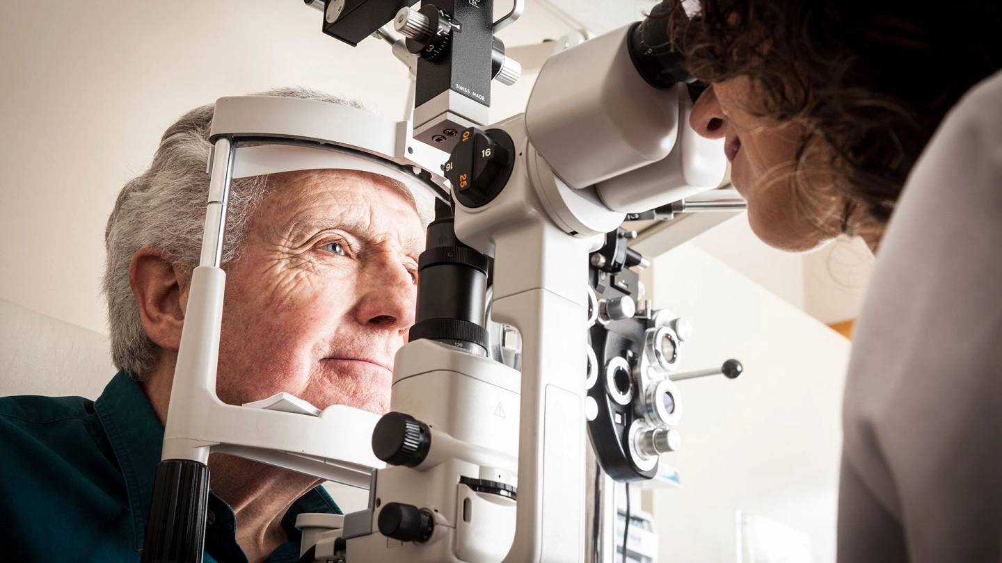
Once the symptoms have been determined and other diseases have been ruled out, the front and middle parts of the eye are examined with a slit lamp (microscope with light).
In order to be able to examine the ocular fundus, the pupils are dilated using special drops. Sight tests determine to what degree central sight has been impacted.
To confirm the diagnosis and define a treatment plan, further tests may be performed in some cases. If wet age-related macular degeneration is suspected, a fluorescein angiography of the retina is performed, for example. A harmless dye is injected in the arm vein, which is then dispersed into the vessels of the retina. A special camera is used to evaluate changes in the eye vessels.
Another test measures retinal thickness with a laser light. The technical term for this is optical coherence tomography (OCT). Among other things, the thickness of the retina is measured with a laser light. This procedure is also used to detect accumulations of fluid. OCT can be used to observe the progression of the disease and adjust treatment as needed.
How is macular degeneration treated?
To date, dry age-related macular degeneration (AMD) can still not be effectively treated.
There are four basic forms of treatment for wet AMD:
- drug treatment
- laser treatment
- surgery
Medication
This is the most important treatment option. VEGF inhibitors are used here. VEGF is a growth factor that promotes the regeneration of blood vessels. It stands for vascular endothelial growth factor. VEGF inhibitors are injected into the eye to prevent the formation of new blood vessels. They cannot cure AMD but they can stop or at least slow its progression. Sometimes, the visual acuity even improves during treatment.
Laser therapy
This procedure uses laser beams to heat and destroy the diseased blood vessels. This type of treatment is possible for only a few people with wet AMD. It is less effective and produces more side effects than treatment with VEGF inhibitors.
Surgery
In certain cases, and if no other treatment is effective, the new blood vessels can also be treated through surgery.
Other options
Even if studies have not yet confirmed the preventive effectiveness of dietary supplements, for example vitamin C, vitamin E, zinc, copper, lutein with zeaxanthin or beta carotene, they can possibly slow down the progression of AMD in people with large drusen when combined with certain medications. These people are at higher risk of visual impairment.
Detailed information, for example on the treatment of AMD, can be found at gesundheitsinformation.de.
- Ba J, Peng RS, Xu D et al. Intravitreal anti-VEGF injections for treating wet age-related macular degeneration: a systematic review and meta-analysis. Drug Des Devel Ther 2015; 9: 5397-5405.
- Berufsverband der Augenärzte Deutschlands (BVA), Deutsche Ophthalmologische Gesellschaft (DOG). Altersabhängige Makuladegeneration AMD (Leitlinie Nr. 21). 2023.
- Chakravarthy U, Evans J, Rosenfeld PJ. Age related macular degeneration. BMJ 2010; 340: c981.
- Evans JR, Lawrenson JG. Antioxidant vitamin and mineral supplements for preventing age-related macular degeneration. Cochrane Database Syst Rev 2017; (7): CD000253.
- Evans JR, Lawrenson JG. Antioxidant vitamin and mineral supplements for slowing the progression of age-related macular degeneration. Cochrane Database Syst Rev 2023; 9(9): CD000254.
- Gao Y, Yu T, Zhang Y, Dang G. Anti-VEGF Monotherapy Versus Photodynamic Therapy and Anti-VEGF Combination Treatment for Neovascular Age-Related Macular Degeneration: A Meta-Analysis. Invest Ophthalmol Vis Sci 2018; 59(10): 4307-4417.
- Lawrenson JG, Evans JR. Omega 3 fatty acids for preventing or slowing the progression of age-related macular degeneration. Cochrane Database Syst Rev 2015; (4): CD010015.
- National Institute for Health and Care Excellence (NICE). Age-related macular degeneration (NICE Guidelines; No. NG82). 2018.
- Solomon SD, Lindsley K, Vedula SS et al. Anti‐vascular endothelial growth factor for neovascular age‐related macular degeneration. Cochrane Database Syst Rev 2019; (3): CD005139.
- Virgili G, Michelessi M, Parodi MB et al. Laser treatment of drusen to prevent progression to advanced age-related macular degeneration. Cochrane Database Syst Rev 2015; (10): CD006537.
- Waugh N, Loveman E, Colquitt J et al. Treatments for dry age-related macular degeneration and Stargardt disease: a systematic review. Health Technol Assess 2018; 22(27): 1-168.
- Wormald R, Evans JR, Smeeth LL, Henshaw KS. Photodynamic therapy for neovascular age-related macular degeneration. Cochrane Database Syst Rev 2007; (3): CD002030.
In cooperation with the Institute for Quality and Efficiency in Health Care (Institut für Qualität und Wirtschaftlichkeit im Gesundheitswesen, IQWiG).
As at:


