Conditions Aortic stenosis
ICD codes: I34 What are ICD codes?
Heart valve diseases cause damage to the heart valves that prevents them from working normally. They frequently occur in older age groups. The most common heart valve disease is aortic stenosis, i.e. a narrowing of the heart valve located between the left ventricle of the heart and the aorta.
At a glance
- Heart valve diseases usually cause damage to the heart valves over many years.
- The most common type of heart valve disease is aortic stenosis (AS).
- With this condition, the valve between the left ventricle of the heart and the aorta becomes narrowed.
- Heart valve diseases are somewhat more common in men than in women.
- If there is only a slight change to a heart valve, it may be sufficient to have this checked regularly by a doctor.
- If the change is causing symptoms or if the disease progresses, various treatment measures are available.
Note: The information in this article cannot and should not replace a medical consultation and must not be used for self-diagnosis or treatment.
")
What is aortic stenosis?
The four valves in the human heart are flaps that function in the same way as mechanical valves by allowing blood to flow in one direction only. They ensure that the heart muscle always pumps blood in the right direction. Oxygen-poor blood is pumped into the lungs, while oxygen-rich blood is pumped back out of the lungs and into the body.
With heart valve diseases, the valves in the heart typically become damaged over the course of a person’s life. Normally, this only occurs at an advanced age due to progressive wear and tear over time. Doctors therefore refer to acquired heart valve disease.
The aortic valve is located between the left ventricle (lower chamber) of the heart and the aorta. If the opening of this valve is narrowed, the heart needs to work harder to pump enough blood into the aorta.
Treatment isn’t always necessary. However, if the damage is causing symptoms or if the patient is at risk of developing complications, doctors usually replace the narrowed valve with a valve prosthesis. This prosthesis can be inserted into the heart during open heart surgery or in a procedure using a catheter.
Video How does the heart function?
This video provides more information about the function and role of the heart.
This and other videos can also be found on YouTube
Watch nowThe privacy policy indicated there applies.
What are the symptoms of aortic stenosis?
People with aortic stenosis don’t usually experience any symptoms until the heart is no longer able to pump enough blood around the body. If a heart valve is only slightly narrowed, the heart can often compensate for the reduced blood flow by means of a stronger heartbeat. As a result, a mild stenosis of the aortic valve may go unnoticed for a long time.
However, if the aortic valve is significantly damaged, blood builds up and collects in the heart, lungs, and, finally, in the circulatory system as a whole.
At this point, symptoms such as the following may occur:
- weakness and reduced performance
- swelling, especially in the lower legs
- irregular, fast, or slow pulse rate
- cough and shortness of breath, especially at night
- chest tightness and chest pain
- fainting
What causes aortic stenosis?
Wear and tear is the main cause of acquired heart valve diseases such as aortic stenosis. Wear and tear in this sense means that the valves in the heart become increasingly calcified with age. In other words, calcium deposits build up on the valves, making them less mobile.
Important: In some people, the heart valves have a different shape from birth. This increases the likelihood that a valve will eventually narrow or fail to close properly. However, the function of the heart valve can also be impaired if the heart muscle is damaged in the peripheral area of a heart valve, e.g. by a heart attack.
How common are heart valve diseases?
Acquired heart valve diseases, such as aortic stenosis due to wear and tear, occur mainly in older people.

About 13% of people aged 75 and older have problems with a heart valve. Heart valve diseases generally affect men somewhat more frequently than women.
What is the outlook for aortic stenosis?
In many people with aortic stenosis, the heart is able to compensate for the narrowing of the aortic valve for a number of years or even decades. However, this means that the heart muscle has to constantly work harder than normal. As a result, the muscle thickens and the heart becomes enlarged.
If the heart is subjected to constant strain, these changes become progressively more pronounced and irreversible.
The thickened heart muscle is no longer as elastic as it used to be. In addition, the ventricles become “worn out”. The heart becomes increasingly weak overall. The resulting discomfort is first manifested only during physical exertion, but later also while at rest. Life-threatening heart failure can be the result.
Can aortic stenosis be prevented?
Smoking increases the risk of cardiovascular disease in general, including heart valve disease. Quitting smoking is therefore one of the most important preventive measures.

An increased risk of heart valve damage can also occur as a result of inflammation of the inner lining of the heart, known as endocarditis. Endocarditis is usually caused by bacteria. For this reason, people with a congenital valve defect or with a new heart valve prosthesis are sometimes given an antibiotic as a preventive measure before having certain dental treatments or medical procedures in their mouths. This kills bacteria that can pass from the mouth into the blood and cause endocarditis.
How is aortic stenosis diagnosed?
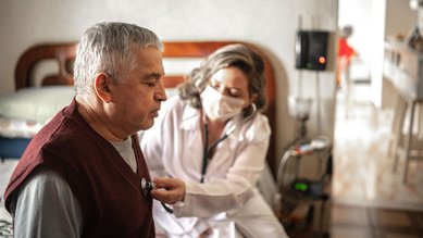
Doctors can obtain important information about whether a heart valve disease is present by performing a thorough physical examination and, in particular, by listening to heart sounds through a stethoscope.
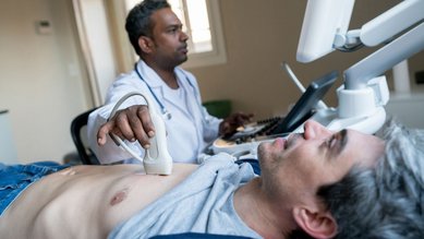
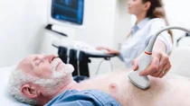
An ultrasound examination of the heart, known as echocardiography, can confirm the diagnosis and reveal how severely the heart valve is damaged. Sometimes an ultrasound examination through the esophagus is useful. In this procedure (known as a transesophageal echocardiography), a tube with an ultrasound probe is inserted up to the level of the heart. This allows for better imaging of the heart compared to a conventional ultrasound scan.
Once heart valve disease has been reliably diagnosed, the doctor also determines the person’s general state of health. Only then is the appropriate treatment selected.
The examinations used for this purpose include, for example:
- resting and stress ECG (electrocardiogram of the heart)
- measurement of blood pressure
- blood sampling for a blood count
- x-ray examination, if necessary computed tomography (CT) and magnetic resonance imaging (MRI)
In addition, an examination with a cardiac catheter may be necessary if there is also a suspicion that the coronary arteries are narrowed. A contrast medium is delivered into the coronary vessels via a cardiac catheter, allowing the doctor to assess the vessels on the screen.
How is aortic stenosis treated?
Slight changes to the aortic valve often don’t require treatment and, in most cases, are simply monitored. If the changes progress and symptoms occur or if a checkup detects signs that the disease is worsening, various therapeutic measures can be considered. Many people with aortic stenosis are given a heart valve prosthesis. However, other treatment options are also available.
Choosing a suitable treatment depends on various factors, such as the age of the patient, the cause and severity of the heart valve disease, and the presence of any other health conditions.
A valve prosthesis can be inserted into the heart during open heart surgery or in a procedure using a catheter. Two different types of prosthesis are used – mechanical and biological.
Mechanical valve prosthesis
This valve prosthesis is made of plastic and metal with mechanical flaps, and its is very durable. It is used in particular for young people, but even then only in rare cases. The reason for this is that there is a risk of blood clots developing on these valves. The blood clots can then travel around the body in the blood stream and cause a blockage in a blood vessel (embolism).
This type of complication can be very serious, and so people who have a mechanical valve prosthesis take anticoagulant medication for the rest of their lives to inhibit the formation of blood clots. Bleeding in the gastrointestinal tract and the brain are potential, though rare, side effects of this medication.
Biological valve prosthesis (bioprosthetic valve)
A bioprosthetic valve may be taken from the heart tissue of a pig or cow, for example. Hearts valves may also be taken from deceased human donors, although this is the exception rather than the rule. The disadvantage of bioprosthetic valves is that they are less durable than mechanical prostheses. From 10 to 15 years after the operation, there is a risk that a biological prosthesis will become calcified and need to be replaced.
Bioprosthetic valves are mostly used in patients over 60 years of age or in younger people who are unable to take anticoagulant medication on a permanent basis. Bioprosthetic valves eliminate the need for long-term medication to inhibit blood clotting.
What is the rehabilitation process after heart valve surgery?
Heart valve surgery is usually followed by a period of rehabilitation (rehab). It helps the patient become accustomed to physical exertion again and gradually increase it. In this way, rehab can help improve the patient’s quality of life. Furthermore, during rehab, medications are adjusted and the heart is checked regularly.
What is it like to live with heart valve disease?
A heart valve disease such as aortic stenosis can also be a psychological burden. Symptoms such as weakness and fatigue can also contribute to an increasingly limited everyday life.
Deciding on a particular treatment can also prove complex and difficult. Questions or uncertainties should therefore be discussed with the attending physician and, if possible, with loved ones.
After heart surgery, it is more difficult to cope with stress for some time. It can therefore be useful to talk to relatives and friends early on about who can help in this phase, when, and how. It is also possible to apply for support from health insurance providers, even before the operation, for example, for help with housework.
- Baumgartner H, Falk V, Bax JJ etc al; ESC Scientific Document Group. 2017 ESC/EACTS Guidelines for the management of valvular heart disease. Eur Heart J. 2017 Sep 21;38(36):2739-2791. doi: 10.1093/eurheartj/ehx391. PMID: 28886619.
- Berg SK, Zwisler AD, Pedersen BD et al. Patient experiences of recovery after heart valve replacement: suffering weakness, struggling to resume normality. BMC Nurs. 2013 Sep 26;12(1):23. doi: 10.1186/1472-6955-12-23. PMID: 24070399; PMCID: PMC3849933.
- Deutsche Herzstiftung. 28. Deutscher Herzbericht 2016. Sektorenübergreifende Versorgungsanalyse zur Kardiologie, Herzchirurgie und Kinderherzmedizin in Deutschland. Deutsche Herzstiftung: Frankfurt am Main 2016.
- Hansen TB, Zwisler AD, Berg SK et al. Cardiac rehabilitation patients' perspectives on the recovery following heart valve surgery: a narrative analysis. J Adv Nurs. 2016 May;72(5):1097-108. doi: 10.1111/jan.12904. Epub 2016 Jan 22. PMID: 26799453.
- Kasper DL, Fauci AS, Hauser SL et al. Harrison's Principles of Internal Medicine. McGraw-Hill: New York 2015.
In cooperation with the Institute for Quality and Efficiency in Health Care (Institut für Qualität und Wirtschaftlichkeit im Gesundheitswesen – IQWiG).
As at: