Conditions Baker’s cysts
ICD codes: M71.2 What are ICD codes?
In adults, Baker’s cysts at the back of the knee are usually caused by damage to the knee joint. They can cause mild pain and feelings of tightness. In most cases, the cysts clear up on their own without treatment.
At a glance
- A Baker’s cyst occurs when excess fluid accumulates at the back of the knee.
- It can cause mild pain and feelings of tightness.
- In adults, the cysts usually form as a result of injury or a chronic joint disease, such as osteoarthritis or rheumatoid arthritis.
- In many cases, smaller cysts clear up on their own without treatment.
- Cooling and stretching the knee may relieve the pain.
- In the event of more severe discomfort, it is important to identify and treat the cause of the cyst.
Note: The information in this article cannot and should not replace a medical consultation and must not be used for self-diagnosis or treatment.
")
What is a Baker’s cyst?
A Baker’s cyst is an accumulation of fluid at the back of the knee. This leads to swelling in the joint capsule area.
The upper and lower leg bones meet at the knee joint. Between them is the narrow joint cavity. To keep the joint working smoothly, the cavity is filled with fluid. The joint and joint cavity are surrounded by the joint capsule, which also contains cushioning sacs of fluid that are called bursae.
In the event of a Baker’s cyst, a build-up of excess joint fluid occurs. This forces its way into the posterior bursa sac, resulting in visible swelling at the back of the knee.
In adults, Baker’s cysts usually occur due to an injury or an inflammation in the knee joint. Depending on the size of the cyst, it may frequently be accompanied by a feeling of tightness or pain in the knee.
Small Baker’s cysts often go unnoticed and clear up on their own without treatment. Treatment is necessary if symptoms develop.
Baker’s cysts are common in people over the age of 50 and in those with knee problems: between 5 and 40 of every 100 people with chronic knee pain have a Baker’s cyst. It is rare for children to have Baker’s cysts.
What are the symptoms of Baker’s cysts?
Larger cysts may cause the following symptoms:
- feelings of stiffness and tightness behind the knee
- knee pain
- stiffness in the knee joint
- swelling at the back of the knee, sometimes visible as a bulge
- swollen calf
The swelling and pain are often worse if the knee is moved a lot.
What causes Baker’s cysts?
In adults, Baker’s cysts often develop after a knee injury, such as a torn meniscus, or as a result of a chronic joint disease, such as rheumatoid arthritis or osteoarthritis.

If the knee is damaged, it is no longer able to sufficiently dampen friction and impacts. To compensate for this, more joint fluid is produced in the joint capsule. This clear, viscous bodily fluid supplies the cartilage in the knee joint with nutrients and acts as a “joint lubricant”. The excess joint fluid forces its way into the bursa sac at the back of the knee, which is connected to the joint cavity. This becomes distended and a Baker’s cyst develops.
If the original knee problem goes away of its own accord, for example acute inflammation in the event of arthritis of the knee, the Baker’s cyst may also go away without treatment.
Which factors increase the risk of developing a Baker’s cyst?
The stability of the knee capsule declines with age. Frequent exertion or excessive strain can cause tiny cracks in the capsule. This, in turn, enables an exchange of fluid between the joint cavity and bursae in the knee capsule – increasing the risk of Baker’s cysts developing.
Older adults are also more likely to have suffered a knee injury at some point in their lives. Chronic joint diseases are also more likely to occur with age. Both of these factors increase the risk of developing a Baker’s cyst.
What are the possible complications of a Baker’s cyst?
If the cause of the Baker’s cyst is treated and less joint fluid is thus formed, the cyst will also go away. However, Baker’s cysts can also remain for years if the underlying condition cannot be effectively treated.
Baker’s cysts can sometimes burst (rupture). In such cases, the joint fluid escapes and disperses into the surrounding tissue, such as the calf muscles. This usually results in sudden, severe pain in the knee and calf. It may also lead to bruising (hematomas). The escaped fluid is then slowly broken down again. Sometimes, the tissue becomes inflamed after the Baker’s cyst ruptures. A visit to the doctor is therefore advisable.
If the cyst presses on blood vessels, a build-up of fluid (edema) can ensue, causing the calf to swell. If the Baker’s cyst presses on nerves, this may cause numbness and muscle weakness in the calf. However, such complications are rare.
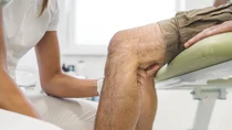
How is a Baker’s cyst diagnosed?
A Baker’s cyst is most clearly visible when the affected leg is stretched out. The doctor can then see and feel the solid bulge where the fluid has accumulated.
When the knee is bent, pressure is removed from the cyst, so it becomes softer or may even disappear entirely.
Further tests are required if the diagnosis is not clear. In these cases, doctors will use imaging techniques, such as:
- ultrasound
- magnetic resonance imaging (MRI)
These techniques make it easier to detect changes to the joints or tissue. Small cysts are also easier to see on an ultrasound scan.
How are Baker’s cysts treated?
Baker’s cysts are only treated if symptoms develop. Potential self-help measures include raising the leg after exercise, bandages or soothing ointments.

Anti-inflammatory pain medication and physiotherapy may also help. The goal of physiotherapy is to keep the knee mobile and to stabilize it. This is achieved through targeted training of the leg muscles.
The knee joint or Baker’s cyst can be punctured in an orthopedic practice. This involves inserting a needle into the cyst or knee joint and extracting the fluid. If severe pain cannot be otherwise alleviated, surgery is sometimes an option – but only if this makes it possible to treat the cause of the Baker’s cyst.
Further information about treating Baker’s cysts can be found at gesundheitsinformation.de.
- Braun J, Müller-Wieland D. Basislehrbuch Innere Medizin. Urban und Fischer: München 2018.
- Brazier BG, Sudekum SA, DeVito PM et al. Arthroscopic Treatment of Popliteal Cysts. Arthrosc Tech. 2018 Oct 8;7(11):e1109-e1114. doi: 10.1016/j.eats.2018.07.006.
- Deutsche Gesellschaft für Orthopädie und Unfallchirurgie e.V. (DGOU). Gonarthrose. S3-Leitlinie. AWMF-Registernummer 187-050. 05.2025.
- Faller A, Schünke M. Der Körper des Menschen. Einführung in Bau und Funktion. Thieme: Stuttgart: 2020.
- Fielding JR, Franklin PD, Kustan J. Popliteal cysts: a reassessment using magnetic resonance imaging. Skeletal Radiol. 1991;20(6):433-5. doi: 10.1007/BF00191086.
- Frush TJ, Noyes FR. Baker's Cyst: Diagnostic and Surgical Considerations. Sports Health. 2015 Jul;7(4):359-65. doi: 10.1177/1941738113520130.
- Han JH, Bae JH, Nha KW et al. Arthroscopic Treatment of Popliteal Cysts with and without Cystectomy: A Systematic Review and Meta-Analysis. Knee Surg Relat Res. 2019 Jun 1;31(2):103-112. doi: 10.5792/ksrr.18.068.
- Hayashi D, Roemer FW, Dhina Z et al. Longitudinal assessment of cyst-like lesions of the knee and their relation to radiographic osteoarthritis and MRI-detected effusion and synovitis in patients with knee pain. Arthritis Res Ther. 2010;12(5):R172. doi: 10.1186/ar3132.
- Herold G. Innere Medizin. Herold: Köln 2021.
- Janzen DL, Peterfy CG, Forbes JR et al. Cystic lesions around the knee joint: MR imaging findings. AJR Am J Roentgenol. 1994 Jul;163(1):155-61. doi: 10.2214/ajr.163.1.8010203.
- Langsfeld M, Matteson B, Johnson W et al. Baker's cysts mimicking the symptoms of deep vein thrombosis: diagnosis with venous duplex scanning. J Vasc Surg. 1997 Apr;25(4):658-62. doi: 10.1016/s0741-5214(97)70292-1.
- Leib AD, Roshan A, Foris LA, Varacallo M. Baker's Cyst. [Updated 2020 July 17]. In: StatPearls [Internet]. Treasure Island (FL). StatPearls Publishing; 2021 Jan-. Aufgerufen am 28.05.2021.
- Li H, Zhang M, Li Y et al. Comparison of clinical outcomes associated with arthroscopic cyst wall preservation or resection in the treatment of popliteal cyst: a systematic review and meta-analysis. Arch Orthop Trauma Surg. 2021 Oct;141(10):1741-1752. doi: 10.1007/s00402-021-03812-4.
- Macfarlane DG, Bacon PA. Popliteal cyst rupture in normal knee joints. Br Med J. 1980 Nov 1;281(6249):1203-4. doi: 10.1136/bmj.281.6249.1203.
- Miller TT, Staron RB, Koenigsberg T et al. MR imaging of Baker cysts: association with internal derangement, effusion, and degenerative arthropathy. Radiology. 1996 Oct;201(1):247-50. doi: 10.1148/radiology.201.1.8816552.
- Neubauer H, Morbach H, Schwarz T et al. Popliteal cysts in paediatric patients: clinical characteristics and imaging features on ultrasound and MRI. Arthritis. 2011;2011:751593. doi: 10.1155/2011/751593.
- Sansone V, de Ponti A, Paluello GM et al. Popliteal cysts and associated disorders of the knee. Critical review with MR imaging. Int Orthop. 1995;19(5):275-9. doi: 10.1007/BF00181107.
- Van Nest DS, Tjoumakaris FP, Smith BJ et al. Popliteal Cysts: A Systematic Review of Nonoperative and Operative Treatment. JBJS Rev. 2020 Mar;8(3):e0139. doi: 10.2106/JBJS.RVW.19.00139.
- Zhou XN, Li B, Wang JS et al. Surgical treatment of popliteal cyst: a systematic review and meta-analysis. J Orthop Surg Res. 2016 Feb 15;11:22. doi: 10.1186/s13018-016-0356-3.
In cooperation with the Institute for Quality and Efficiency in Health Care (Institut für Qualität und Wirtschaftlichkeit im Gesundheitswesen – IQWiG).
As at: