Conditions Dunbar syndrome
ICD codes: I77.4 What are ICD codes?
Dunbar syndrome is rare. If it causes symptoms, the most typical symptom is recurring pains in the stomach. This happens because a large blood vessel in the abdomen becomes compressed by a band of connective tissue.
At a glance
- Dunbar syndrome occurs when a band of connective tissue constricts a large blood vessel that supplies blood to the organs in the upper abdomen.
- It is a rare condition that occurs more frequently in women than in men.
- In most cases, compression of the blood vessel causes no problems but some people experience symptoms.
- Stomach ache is typical, and often occurs after eating and sometimes after physical movement. However, the pain may also be constant.
- To diagnose the syndrome, specific techniques are used to view the blood vessels and blood flow.
- Surgery is the only treatment for this condition.
Note: The information in this article cannot and should not replace a medical consultation and must not be used for self-diagnosis or treatment.

")
What is Dunbar syndrome?
The celiac artery (also called the celiac trunk or truncus coeliacus) is a large blood vessel that supplies blood to the organs in the upper abdomen. The median arcuate ligament of the diaphragm – a band of connective tissue in the lower part of the chest – is located close to this blood vessel. If this ligament presses on the artery and causes discomfort, this is known as Dunbar syndrome. Other names for this condition are median arcuate ligament syndrome (MALS), celiac artery compression syndrome (CACS), celiac axis syndrome, truncus coeliacus compression syndrome and Harjola-Marable syndrome.
What are the symptoms of Dunbar syndrome?
Stomach pains are a typical symptom of Dunbar syndrome. The pains may vary in their intensity and usually occur after eating. For some people, exercise and movement can also trigger pain. There are also others who experience this symptom while resting, on a recurring basis or even constantly.
Other symptoms may also occur, such as:
- weight loss
- nausea
- vomiting
- diarrhea
- abnormal sounds from the stomach
- pressure pain in the upper abdomen
- delayed emptying of the stomach
What causes Dunbar syndrome?
The median arcuate ligament is a strong band of connective tissue that connects the right and left sides of the diaphragm. This ligament forms the front of the opening in the diaphragm, through which the aorta passes, for example.
Below this band of tissue, a large blood vessel called the celiac artery branches off from the aorta. This artery supplies blood to organs including the stomach, liver and small intestine.
The exact position of the ligament and blood vessel varies from person to person. In some people, the ligament is too low in the chest. In rarer cases, the celiac artery branches off from the aorta at a higher point than normal. Both of these deviations can cause the ligament to compress the artery.
Most people don’t notice any effects as a result because other arteries also supply blood to the organs in the abdomen. It is unclear why other people experience problems but it is suspected that there is another cause at play that is connected with a group of nerve cells called the celiac ganglion, located close to the celiac artery.
It forms part of an important network of nerves. It may be that the ligament sometimes also compresses this ganglion, contributing to the pain experienced in Dunbar syndrome.
How common is Dunbar syndrome?
Dunbar syndrome is rare. It occurs more frequently in women than in men, between the ages of 40 and 60 and in people with a slight frame.
The condition can also occur in children.

What is the outlook for Dunbar syndrome?
Recurring pressure on the artery causes it to change over time. The wall of the artery thickens and the blood vessel may become partly or fully blocked.
Below the narrowed portion, the artery may expand and bulge outward, forming an aneurysm.
How is Dunbar syndrome diagnosed?
The symptoms that are typical of Dunbar syndrome also occur in several other diseases of the stomach, bowel, gall bladder and liver.
As Dunbar syndrome often causes no symptoms, doctors normally run a series of tests to exclude other diseases first.
These tests may include extensive blood tests, an ultrasound scan of the liver, pancreas and gallbladder, as well as an endoscopy/gastroscopy to examine the stomach or colonoscopy to examine the bowel.
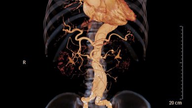
Various imaging techniques are also used to visualize the blood vessels and blood flow.
Interesting fact: A Doppler ultrasound scan – together with a computed tomography (CT) scan or magnetic resonance imaging (MRI) scan – usually provides reliable results for diagnosis.
Another useful diagnostic tool is a nerve block. This involves administering a local anesthetic to the nerves in the celiac ganglion while using CT monitoring to determine whether the pain is reduced.
How is Dunbar syndrome treated?
Symptoms caused by Dunbar syndrome require surgical intervention, as they cannot be relieved by medication.

During the procedure, the surgeon divides the ligament of the diaphragm to relieve the pressure on the artery. The celiac ganglion is also frequently removed, as this group of nerve cells is thought to play a role in causing Dunbar syndrome.
Where possible, the procedure is performed using an endoscope, involving only small incisions in the abdominal wall. An endoscope is a fine tube with a light source and camera attached to it. It enables monitoring of the surgical site throughout the procedure. Small instruments can also be inserted and used through the tube. This type of procedure is also known as laparoscopic surgery or minimally invasive surgery.
Most people experience an immediate improvement in symptoms following surgery. In a small number of patients, the procedure provides no relief or the pains quickly return.
It may also take several months for the symptoms to resolve. In addition, symptoms may disappear only to recur again after an extended period of time – this happens in almost 10 percent of people who experience relief following surgery.
What is the rehabilitation process after surgery for Dunbar syndrome?
Most patients need to stay in hospital for 2 to 3 days following laparoscopic surgery. The recovery period is usually short because the procedure only requires tiny incisions in the abdominal wall and so the wound is small.
- Goodall R, Langridge B, Onida S et al. Median arcuate ligament syndrome. Journal of Vascular Surgery 2029; Volume 71. Number 6, S.2170-2176. doi: 10.1016/j.jvs.2019.11.012. Epub 2019 Dec 25. PMID: 31882314.
- Saleem T, Katta S, Baril DT. Celiac Artery Compression Syndrome. [Updated 2021 Aug 13]. In: StatPearls [Internet]. Treasure Island (FL): StatPearls Publishing; 2021 Jan-. Aufgerufen am 13.04.2021.
- UpToDate (Internet). Celiac artery compression syndrome. Wolters Kluwer 2021. Aufgerufen am 13.04.2021.
In cooperation with the Institute for Quality and Efficiency in Health Care (Institut für Qualität und Wirtschaftlichkeit im Gesundheitswesen – IQWiG).
As at: