Conditions Surfer’s eye (pterygium)
ICD codes: H11.0 What are ICD codes?
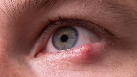
Surfer’s eye is a growth of tissue that develops on the conjunctiva, i.e. the clear film of tissue covering the white of the eye. It is often visible as a patch or area of cloudy white. In most cases, surfer’s eye causes no symptoms, although it may cause distress from a cosmetic point of view. In some cases, it can irritate the eye or affect vision.
At a glance
- Surfer’s eye is often visible in the eye as a triangular patch of tissue on the eye.
- It is thought that the condition is caused by prolonged exposure to strong sunlight.
- In most cases, surfer’s eye causes no symptoms. However, people may find it distressing from a cosmetic point of view.
- In some cases, the tissue growth may also lead to visual impairment or an astigmatism (an irregularity in the curvature of the eye).
- If surfer’s eye causes symptoms, it can be treated with lubricating eye-drops. If vision is affected, surgical removal may be considered.
- It’s important to protect the eyes from strong UV radiation in sunlight – wearing sunglasses and a hat could prevent surfer’s eye.
Note: The information in this article cannot and should not replace a medical consultation and must not be used for self-diagnosis or treatment.

What is surfer’s eye?
The condition is known as “surfer’s eye” because it occurs in people who spend a lot of time outdoors in bright sunlight. A growth of this type is also known as a “pterygium”, which comes from the Greek word for “wing” because the tissue growth can be described as triangular and wing-like in shape – similar to the wing of an insect.
The exact cause is unknown. However, surfer’s eye occurs most commonly in southern and tropical regions of the Earth, and so doctors suspect that it is connected with exposure to ultraviolet radiation (UV light).
People who develop surfer’s eye often spend a lot of time in bright sunlight, wind or a dusty atmosphere.
If there are no symptoms, the growth must simply be monitored. However, if it causes itchy, irritated eyes, it may need to be treated with special eye drops and medication.
If vision is affected, the pterygium may be surgically removed. Surgery may also be performed if the patient finds the condition distressing from a cosmetic point of view.
It is estimated that around 1.6 million people in Germany have surfer’s eye.
What are the symptoms of surfer’s eye?
The tissue growth initially appears as a thickening of the skin, a patch of skin or a flap of skin in the inner corner of the eye next to the nose, with a shape somewhat similar to the triangular shape of an insect wing. However, the growth may also appear as a small spot or patch or as a whitish area of cloudiness.
Sometimes, the pterygium extends from the conjunctiva onto the cornea (the membrane covering the front part of the eye near the pupil), where it is visible as a cloudy white patch on the iris (the colored portion of the eye).
In most cases, surfer’s eye causes no symptoms. However, some people are bothered by the condition, as it makes them feel as if they have something stuck in their eye and can make it more difficult to put in a contact lens, for example. It may feel as though a fly or dust particle is trapped in the eye.
The eye may also feel dry and irritated and appear red. Scratching or rubbing makes the irritation worse and may cause inflammation.
In rare cases, the growth is severe enough to impair vision and affect the ability of the eye to focus.
What causes surfer’s eye?
The exact cause of surfer’s eye is not yet known. While several factors seem to increase the risk of this tissue growth developing, the most important of these is exposure to sunlight. Surfer’s eye is particularly likely to occur in people who spend a lot of time in bright sunlight.
This is why the condition is much more common in regions with extremely high levels of UV radiation. It is suspected that strong sunlight activates growth factors in the cells, which in turn leads to tissue growth on the eye.
More recent research also indicates a connection between surfer’s eye and infection with human papillomaviruses (HPV). These viruses may be responsible for triggering tissue growth. For example, some virus types cause warts, while other lead to tissue changes in the neck of the womb (cervix).
Surfer’s eye is more common among people who have previously been infected with HPV.
How does surfer’s eye develop?
Most people with surfer’s eye have no symptoms and don’t require any treatment.
However, surfer’s eye sometimes gets worse over time. As the growth impairs the production of tears to protect the eyes, it may cause irritations and redness once it reaches an advanced stage.
If the growth reaches the cornea, it can cause vision impairments. The tissue growth may also change the curvature of the cornea. This can result in astigmatism – where the cornea is so curved that vision becomes blurred and distorted.
How can surfer’s eye be prevented?
As strong sunlight may increase the risk of developing surfer’s eye, it is advisable to protect the eyes from prolonged exposure to high levels of UV radiation.
Sunglasses and a wide-brimmed hat or visored cap shield and protect the eyes. Sunglasses with wide or wrap-around levels also prevent eye exposure to sunlight from the side. Strong sun protection for the eyes is particularly important in regions with high levels of UV radiation.
How is surfer’s eye diagnosed?
Doctors can recognize surfer’s eye by means of a visual examination. The wing-like structure of the growth is so distinctive that the condition can normally be diagnosed immediately. An more in-depth examination by an eye specialist can determine the extent of the growth. A sight test determines whether the pterygium is causing a vision impairment.
How is surfer’s eye treated?
The treatment of surfer’s eye depends on whether the vision is impaired and whether the patient is bothered by the tissue growth.
If there are no symptoms such as itching or problems with vision, it is usually sufficient to have the condition checked regularly by a doctor.
Symptoms can be relieved by means of external treatment with lubricating eye-drops, for example in the form of artificial tears. These treatments are used if the growth is not affecting the patient’s sight or movement of their eye.
It is normally sufficient to apply 1 to 2 drops to the affected area between three and four times daily.
If a patient reacts to preservatives in the drops, preservative-free eye-drops are also available.
In addition, anti-inflammatory glucocorticoids or non-steroidal anti-inflammatory drugs (NSAIDs) can be used to treat the symptoms. However, use of these drugs is associated with serious side-effects, such as inflammation of the eye, glaucoma and cataracts.
With severe vision impairments, a pterygium can be surgically removed. However, there is a high probability of it growing back again at a later stage.
- Sarkar P, Tripathy K. Pterygium. [Updated 2021 Aug 21]. In: StatPearls [Internet]. Treasure Island (FL): StatPearls Publishing. 2021 Jan-. Aufgerufen am 02.12.2021.
- DynaMed (Internet), Ipswich (MA). Pterygium. EBSCO Information Services. Record No.T920562. 2018 (1995). Aufgerufen am 02.12.2021.
- UpToDate (Internet). Pterygium. Wolters Kluwer 2020. Aufgerufen am 02.12.2021.
- The College of Optometrists (Internet). Pterygium. Aufgerufen am 02.12.2021.
In cooperation with the Institute for Quality and Efficiency in Health Care (Institut für Qualität und Wirtschaftlichkeit im Gesundheitswesen – IQWiG).
As at:


