Conditions Salivary stones
ICD codes: K11.5 What are ICD codes?
Salivary stones may block the salivary flow, causing a painful swelling in the salivary gland. Symptoms are felt in particular when eating. There are various ways to remove salivary stones.
At a glance
- People have six large salivary glands in the head that produce saliva. They release the saliva into the oral cavity via excretory ducts.
- If an excretory duct is blocked by a salivary stone, the affected gland can become swollen.
- The swelling is particularly likely to occur when eating and can sometimes be painful.
- The swelling may be temporary, recur after a period of time or grow continuously.
- Salivary stones are often flushed out when the salivary flow is stimulated by massaging the salivary glands.
- Surgery is sometimes required to remove the salivary stone.
Note: The information in this article cannot and should not replace a medical consultation and must not be used for self-diagnosis or treatment.

")
What are salivary stones?
Salivary stones are small, calcified masses of saliva. These are deposited in the excretory ducts of the salivary glands, which they can then block.
If this prevents the saliva from flowing freely, the affected gland can become painfully swollen.
People have three large salivary glands on each side of the head:
- the parotid gland
- the submandibular gland (also known as the submaxilliary gland)
- the sublingual gland below the tongue

Salivary stones occur most commonly in the salivary gland located below the jaw bone (the submandibular gland) and usually on one side only. Salivary stones may occur in several places within excretory ducts of the affected gland.
Measures to stimulate salivary flow often help to flush out the salivary stone. In some cases, stones have to be surgically removed.
What are the symptoms of salivary stones?
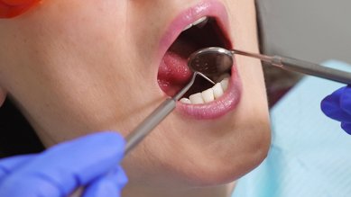
It is sometimes possible to see salivary stones by looking into the oral cavity. They have a recognizable round or oval shape and are white to yellowish in color. Even if they cannot be seen, they can often be felt.
Smaller salivary stones frequently cause no symptoms. However, if a stone grows larger and becomes lodged in the excretory duct, it prevents the saliva from flowing freely out of the duct, causing it to accumulate and flow back into the gland. The salivary gland can then swell and cause pain. The symptoms typically occur when the individual is eating, as this is when saliva production is stimulated.
The salivary glands usually swell repeatedly, at intervals of several days or even weeks. In general, only one side of the face is affected.
If a salivary stone blocks an excretory duct and prevents the saliva from flowing freely, the affected salivary gland can become inflamed and infected with bacteria. Typical symptoms of inflammation include severe pain, redness, swelling and a high temperature.
What causes salivary stones?
The role of the salivary glands is to produce saliva, which is important for swallowing and digestion, among other processes. Saliva leaves the gland and enters the oral cavity through an excretory duct.
This duct may become narrower naturally or due to an inflammation or injury. This facilitates an accumulation of saliva, which in turn may lead to the formation of salivary stones.
The composition of the saliva also has a role to play. Saliva can be watery at some times and have a slimy/mucoid consistency at others, and it contains a wide range of minerals and proteins. It not yet known what causes liquid saliva to form calcified stones.
Tiny calcifications or bacteria may sometimes trigger salivary stones. If the flow of saliva is thick and slow, more and more layers can become deposited on the calcified area. This makes the salivary stone bigger until it ultimately blocks the excretory duct. The saliva then accumulates in the glandular tissue.
Salivary stones are particularly likely to occur in the submandibular glands below the floor of the mouth. This may be because these glands produce a thicker, more mucous and calciferous consistency of saliva than the other salivary glands. In addition, the excretory ducts from these glands are very long and the saliva has to be transported upwards against gravity. As a result, it flows more slowly.
Which factors increase the risk of developing salivary stones?
If people drink too little or take dehydration tablets (diuretics), the body as a whole may contain too little water.
As a result, it produces less saliva, the salivary flow is reduced and the probability of salivary stones increases.
There are also some types of medication that directly affect salivary flow and typically cause a dry mouth.
Various factors can increase a person’s risk of salivary stones:
How common are salivary stones?
Less than one percent of the population will develop a salivary stone that causes symptoms over the course of their life.

Most people who develop a salivary stone are between the ages of 30 and 60. It is very rare for children to develop salivary stones.
3 out of every 4 people who develop salivary stones develop a single stone. Salivary stones affect both sides of the face equally. It is rare for both sides to be affected at the same time.
What possible complications can occur with salivary stones?
When saliva becomes blocked in a salivary gland, the glandular tissue may become inflamed. In most cases, a salivary gland inflammation can be successfully treated with antibiotics. Despite this, pus can accumulate in rare cases. This kind of accumulation of pus is also known as an abscess.
If the salivary gland remains inflamed for a long time, the affected gland can stop working properly and permanently produce less saliva. If the cause of the blockage is eliminated early enough, however, the salivary gland often recovers.
How are salivary stones diagnosed?
Doctors begin by examining the oral cavity and feeling the salivary glands and excretory ducts. They often do this by palpitating the neck from the outside. Doctors can usually successfully detect and locate salivary stones and the associated enlargement of the excretory duct using an ultrasound scan.
In some cases, remedies intended to stimulate the flow of saliva are used during the examination. These can be administered in the form of lozenges, for example. A magnetic resonance imaging (MRI) scan or computed tomography (CT) scan may also be required.
Doctors can also use very narrow medical instruments to look directly into the excretory ducts. This procedure is known as a salivary duct endoscopy (sialoendoscopy). The benefit of this type of examination is that treatment can be administered at the same time. For example, the stone can sometimes be broken up and removed during the salivary duct endoscopy or the salivary duct can be flushed or expanded.
How are salivary stones treated?
Treatment depends on the location and size of the stone as well as the symptoms that it is causing.
Salivary stones that are discovered by accident and are not causing any problems can initially be left untreated.

If the salivary stone is detected for the first time in a patient and is causing problems, the following treatment options are considered first:
- Substances that stimulate the saliva production, such as sour sweets or chewing gum, can serve to flush out salivary stones with the increased salivary flow.
- It is helpful to consider whether the patient is drinking sufficient fluids to facilitate the production of saliva.
- In addition, doctors can enlarge the opening of the excretory duct slightly and massage the salivary gland in the direction of the excretory duct to help loosen the stone.
- If there are signs of a bacterial infection of the salivary gland, antibiotics and anti-inflammatory painkillers are used to address this.
In many people, the symptoms will then recede.
If salivary stones continue to block the exit of an excretory duct, the following further treatment options are available:
- Using a very thin instrument called a micro-endoscope, doctors can locate a salivary stone directly and remove it. They sometimes also make a small incision during this procedure to extend the opening of the excretory duct.
- Shock waves are sometimes used to break salivary stones into small pieces by exposing them to short bursts of pressure waves. The shock waves can be delivered from outside the body or from within the excretory duct itself using an endoscope.
- Some people require surgery using an external incision or through the mouth. Doctors always try first to fully restore the salivary gland and the excretory duct. It is only in rare cases that the entire salivary gland has to be removed.
- Deutsche Gesellschaft für Hals-Nasen-Ohren-Heilkunde, Kopf- und Halschirurgie e.V. (DGHNO-KHC). S2k-Leitlinie. Obstruktive Sialadentitis. 04/2020. AWMF-Registernummer 017-025. Aufgerufen am 29.01.2025.
- DynaMed [Internet]. Ipswich (MA). Salivary Gland Stone. EBSCO Information Services. Aufgerufen am 29.01.2025.
- Hammett JT, Walker C. Sialolithiasis. [Updated 2024 September 10]. In: StatPearls (Internet). Treasure Island (FL): StatPearls Publishing. 2025 Jan-. Aufgerufen am 29.01.2025.
- Badash I, Raskin J, Pei M, Soldatova L, Rassekh C. Contemporary Review of Submandibular Gland Sialolithiasis and Surgical Management Options. Cureus 2022: 14(8);e28147.